Otoacoustic Emissions
An objective test of cochlear function.
DPOAEs or Otoacoustic emissions
Otoacoustic emissions (OAEs) are sounds measured in the external ear canal that are associated with mechanical activity of the outer hair cells in response to sound entering the cochlea. There are two types of OAE used in clinical audiology.
- Distortion Product Otoacoustic Emissions (DPOAE)
- Transient Evoked Otoacoustic Emissions (TEOAE)
In our clinic, we use DPOAE rather than TEOAE, however TEOAE is still widely used in audiology. Alberta Health runs a newborn hearing screening program and uses an automated TEOAE system to provide a Pass/Refer result for neonates as a basic screen of audiological function.
Explaining the fundamental differences between TEOAE and DPOAE is beyond the scope of this guide, but more can be read here.
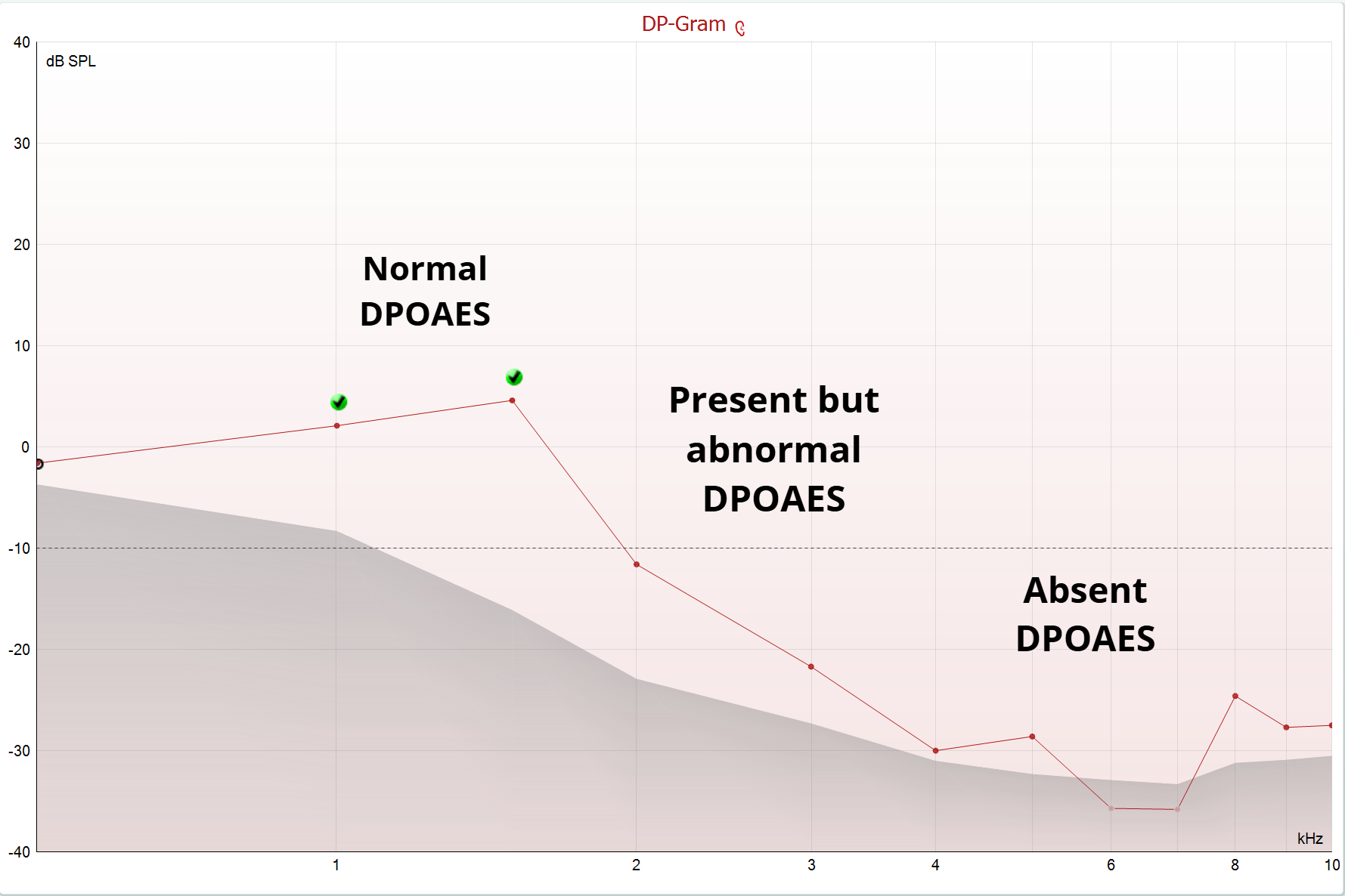
OAES are a very powerful and yet seldomly used clinical tool in adult audiology. Normative data is used to interpret OAE results. OAES tell audiologists little about the degree of hearing loss, but are excellent at identifying which frequencies are likely more affected than others, and they are excellent at identifying normal cochlear function.
We feel that OAES are valuable for:
- Cross checking other test results.
- For example, a dysfunctional cochlea will often have abnormal OAES, often in a pattern that mirrors the frequency ranges being affected by measurable hearing loss.
Ototoxicity screenings to that may occur with certain therapeutics, such as chemotherapy or aminoglycoside antibiotics. - They can help differentiate between retrocochlear and cochlear hearing loss
- Can help monitor for the first signs of noise induced hearing loss.
- Explaining mild difficulties in hearing, misunderstanding, or difficulties limited to noisier situations by identifying areas of reduced motility in the cochlea, without significant hearing loss being present for pure tones.
- OAES can help identify isolated areas of auditory dysfunction that may be related to the perception of tinnitus.
Clinical examples
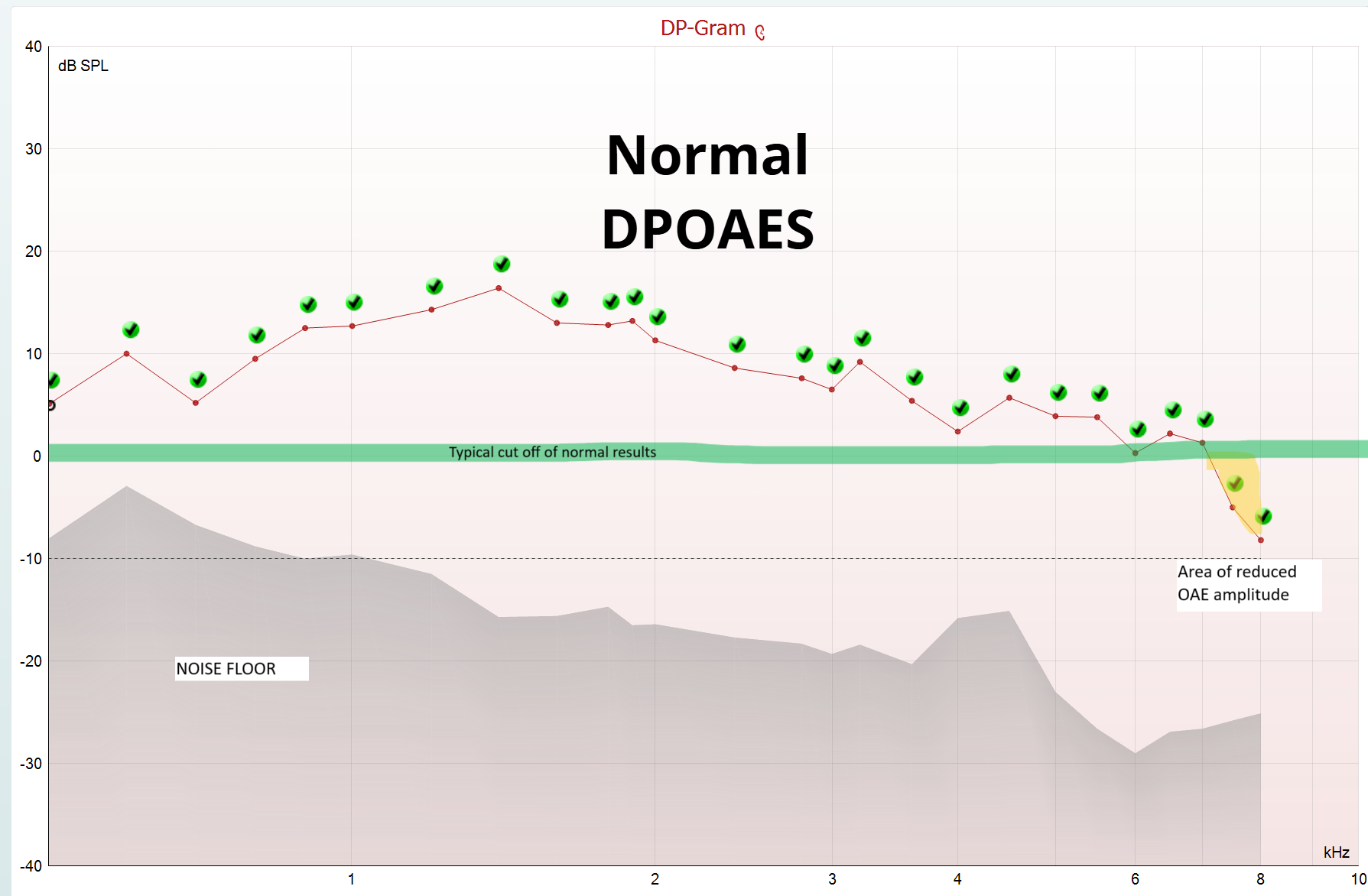
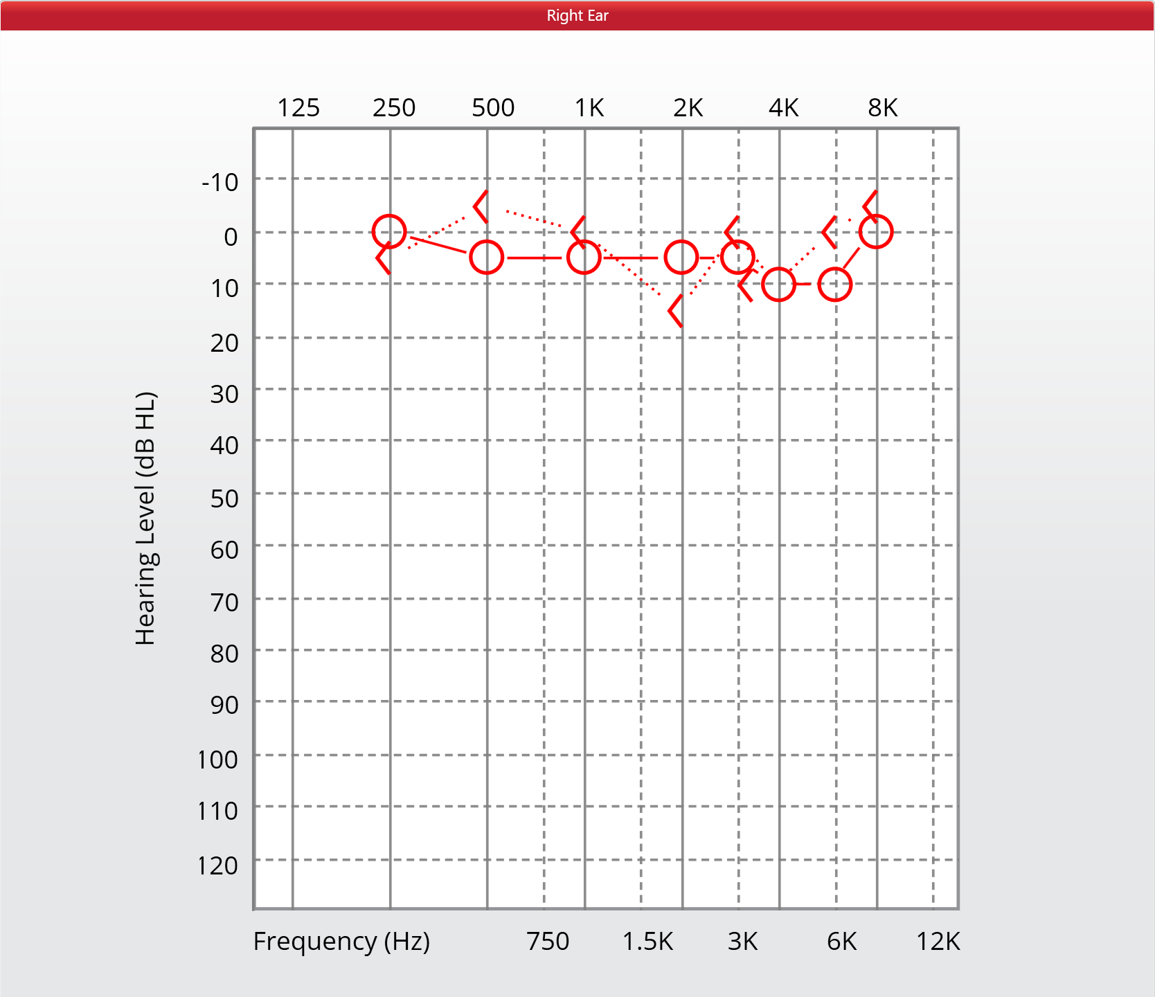
Normal Audiogram, almost normal DPOAE
A Right Sided DPOAE result showing mostly normal function. Note there is a small area of abnormality at 7-8Khz. Hovering over the image will show the audiogram for this patient. Note that the area of abnormality does not appear in the pure tone audiogram. There may be some small areas of damage to the highest frequency regions of the cochlea, or waning sensitivity, but no true hearing loss.
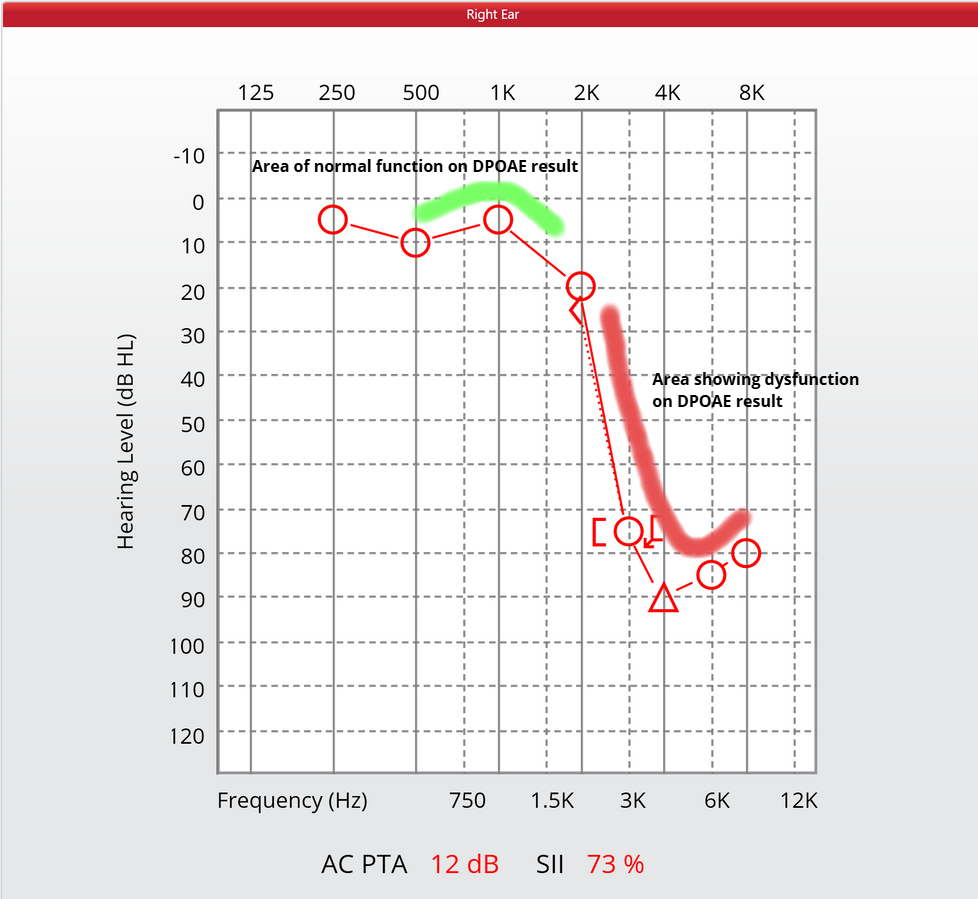
Steeply sloping audiogram, abnormal DPOAE results
An older patient with a steeply sloping audiogram. Note that DPOAEs are still measurable at 1-1.5Khz, which corresponds with the better hearing levels of the audiogram (hover over to see the audiogram)